

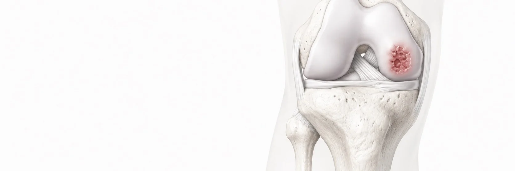
What is osteochondritis dissecans?
Osteochondritis dissecans (OCD) is a condition in which a fragment of cartilage along with the underlying bone gradually separates from the surrounding healthy tissue. It most often affects the medial femoral condyle of the knee in adolescents and young adults, though it can occur in other parts of the joint and at any age.
OCD is one of the great mimickers of knee pain in young athletes, easily missed unless specifically considered. Early diagnosis dramatically changes the treatment options and the prognosis.
What causes OCD?
The exact cause of osteochondritis dissecans is not fully understood. Most specialists think it results from a combination of factors that weaken a small area of bone just beneath the cartilage, most often in the knee of an active child or adolescent.
- Repetitive microtrauma from sport. Repeated small impacts, for example from running and jumping, are thought to stress the bone under the joint surface faster than it can repair.
- Rapid skeletal growth in adolescence. The condition is most common while the skeleton is still growing, when the bone beneath the cartilage is more vulnerable.
- Vascular disturbance. A reduced blood supply to a small area of subchondral bone can prevent it from healing normally.
- Genetic predisposition. OCD occasionally runs in families, and in some patients it appears in both knees.
What are the symptoms?
- vague, activity-related knee pain
- swelling, especially after sport
- stiffness
- in advanced cases, mechanical symptoms, clicking, catching, locking, when the fragment becomes loose
- occasionally, true giving way episodes
How is the diagnosis made?
- clinical examination
- X-rays, including specific tunnel views, often show the OCD lesion clearly
- MRI, the most important investigation, showing the size, location, depth and stability of the fragment, plus the state of the cartilage and any surrounding bone oedema
I always tell patients that early diagnosis is essential. The earlier OCD is identified, particularly in skeletally immature patients, the greater the chance of healing without surgery.
What is the conservative treatment?
In skeletally immature patients with stable OCD lesions, the lesion has an excellent capacity to heal with the right protection:
- modified weight-bearing for several weeks to months, sometimes with bracing
- avoidance of sport and impact loading during healing
- structured physiotherapy to maintain muscle strength and joint mobility
- regular MRI follow-up to monitor healing
Conservative treatment can take 3–6 months and requires patience and compliance, but in well-selected cases delivers excellent results.
When is surgery recommended?
Surgery is recommended for:
- unstable OCD lesions
- loose fragments inside the joint
- skeletally mature patients (closed growth plates) where conservative treatment is less likely to succeed
- lesions that have not healed despite an adequate conservative trial
- persistent symptoms with mechanical features
How is surgical treatment performed?
Surgical treatment depends on the size, location and stability of the lesion. It is performed arthroscopically through small incisions and may include:
- fixation of the fragment with bioabsorbable screws or pins when the fragment is salvageable
- drilling of the lesion to stimulate healing in stable but symptomatic lesions
- removal of unsalvageable loose fragments combined with cartilage restoration techniques (such as the Minced Cartilage technique) for the resulting defect
- in larger or more complex lesions, advanced cartilage restoration procedures
I draw on my training at one of the largest cartilage centres in Europe and apply modern arthroscopic techniques aiming for stable repair and optimal functional recovery.
What should the patient expect after surgery?
- protected weight-bearing for 6–12 weeks, depending on what was performed
- structured rehabilitation under physiotherapist guidance
- return to sport typically at 6–9 months, depending on the procedure and how the cartilage heals