

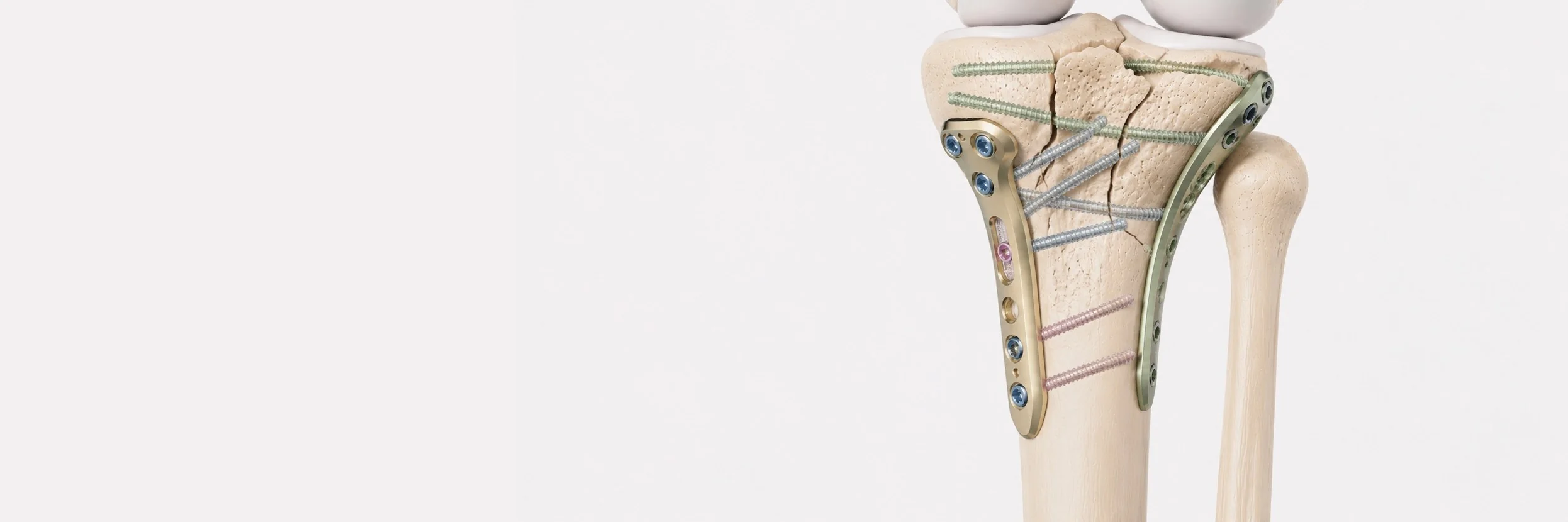
What is the 360° approach to tibial plateau fractures?
The tibial plateau is the upper end of the tibia where it forms the lower part of the knee joint. Fractures here are challenging because the joint surface must be reconstructed as accurately as possible, any residual step-off or incongruity can translate into accelerated long-term joint degeneration.
Modern understanding divides the tibial plateau into three columns: medial, lateral and posterior. Most fracture patterns involve only the anterior columns and can be addressed through a single anterior or anterolateral incision. But fractures involving the posterior column, now increasingly recognised as a distinct fracture pattern, require posterior access, which a single anterior approach cannot provide.
The 360° approach combines both anterior and posterior incisions in a single operation, providing complete visualisation and direct anatomical reduction of all three columns. It is a demanding technique that requires specific training and instrumentation, and offers the best possible opportunity of restoring the joint surface anatomically.
When is the 360° approach needed?
The 360° approach is reserved for specific fracture patterns:
- three-column fractures (Schatzker VI with posterior involvement, or fractures classified as Luo three-column)
- isolated posterior column fractures that cannot be adequately reduced through an anterior approach
- fractures with significant posterior depression that may be missed or insufficiently reduced from an anterior approach alone
- failed previous reduction with persistent posterior displacement
How is the operation planned?
Tibial plateau fractures are unforgiving, meticulous preoperative planning is essential:
- CT scan with 3D reconstruction is mandatory for complex patterns, allowing precise fracture classification according to column involvement
- assessment of the soft tissues, high-energy injuries often have skin and muscle compromise that often require time to recover before definitive fixation
- staging plan, sometimes a temporary external fixator is applied first to maintain limb length and alignment while the soft tissues recover
- careful patient positioning, typically a prone-to-supine combination, repositioning the patient between surgical approaches for the 360° access
- pre-positioned implants, locking plates and screws specifically designed for each column
How is the operation performed?
For the 360° approach, the operation typically proceeds in stages during the same anaesthetic session:
- patient positioning to allow access to both the front and back of the knee
- posterior approach first (often via the postero-medial or postero-lateral window), direct visualisation, reduction and temporary fixation of the posterior column
- patient repositioning to supine
- anterior or anterolateral approach, reduction of the medial and lateral columns, elevation of depressed articular fragments, and definitive fixation with locking plates
- arthroscopy-assisted assessment, the arthroscope is used to confirm anatomical reduction of the joint surface under direct vision, and to address any associated meniscal or cartilage injury that might otherwise remain unrecognised
- bone graft or substitute placed under elevated articular fragments to support the reconstructed joint surface
- final fluoroscopic confirmation of anatomical reduction in all planes
What should the patient expect after surgery?
- hospital stay of 4–7 days, depending on soft-tissue recovery and pain control
- non-weight-bearing on the operated leg for 8–12 weeks while the bone heals
- physiotherapy from day one, focused on restoring early knee motion, postoperative stiffness is one of the most common avoidable complications
- continuous passive motion (CPM) therapy in the early post-operative period in selected cases
- progressive partial weight-bearing from 8–10 weeks depending on radiographic evidence of healing
- full weight-bearing at approximately 12–16 weeks
- driving usually resumes at 8–12 weeks
- return to demanding activities at 6–9 months
How important is rehabilitation?
Rehabilitation is at least as important as the operation. The two major threats to a good outcome are:
- loss of motion (arthrofibrosis), prevented by early, gentle, progressive physiotherapy
- premature weight-bearing, which can lead to collapse of the reconstructed joint surface
A close working relationship with a skilled physiotherapist is essential. I follow patients closely throughout the rehabilitation process, with regular review and X-rays to confirm bone healing before each progression in weight-bearing.
What is the long-term outcome?
Outcomes after well-executed tibial plateau surgery are generally good, but they depend heavily on the accuracy of joint surface reconstruction:
- most patients regain near-normal knee function and mobility when anatomic reduction is achieved
- long-term arthritis risk is reduced (but not eliminated) by accurate reconstruction, even small residual incongruities can lead to accelerated joint wear over years
- return to low-impact sport is usual at 6–9 months
- return to high-impact sport is more variable and depends on the specific fracture