What does hip fracture surgery involve?
Hip fracture surgery is the urgent surgical treatment of fractures of the proximal femur. The choice of operation depends primarily on the location and pattern of the fracture:
- intracapsular (femoral neck) fractures, usually require replacement (hemiarthroplasty or total hip arthroplasty) because of the significant risk of avascular necrosis and non-union
- extracapsular (intertrochanteric and subtrochanteric) fractures, most commonly treated with internal fixation using an intramedullary nail or sliding hip screw
In all cases, the goal is to allow immediate weight-bearing after surgery, mobilising elderly patients as quickly as possible is one of the most important factors in survival and recovery.
Why is timing so important?
Hip fracture surgery is time-sensitive. The evidence is clear: surgery within 24–48 hours of injury is associated with significantly lower complication and mortality rates compared to delayed surgery.
The reason is straightforward: every day a patient spends in bed before surgery brings increased risk of pneumonia, pressure sores, blood clots, urinary infection, and muscle wasting. The operation enables mobilisation, and mobilisation is fundamental to recovery.
How is internal fixation performed?
For extracapsular fractures, I perform internal fixation using minimally invasive surgical approaches:
- closed reduction of the fracture on a a fracture table under fluoroscopic guidance
- insertion of an intramedullary nail through a small incision at the tip of the greater trochanter, with cephalomedullary screws stabilising the fracture into the femoral head
- or, for selected stable fracture patterns, a sliding hip screw with side plate fixation through a slightly larger incision
- final fluoroscopic confirmation of reduction and implant position
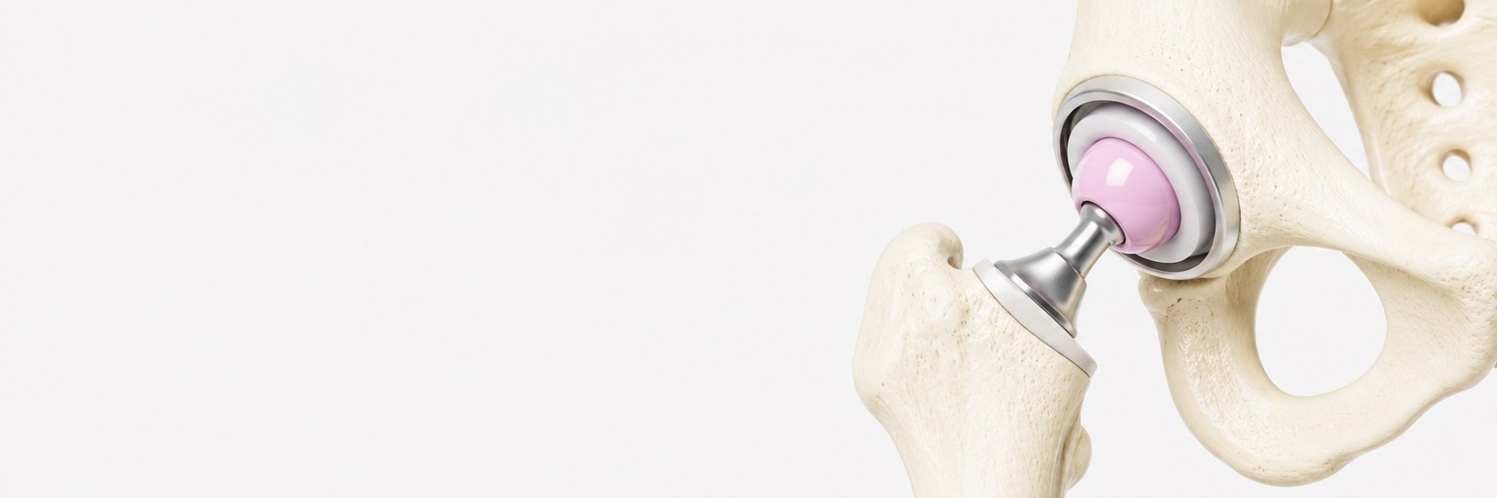
How is hip replacement performed for fracture?
For displaced intracapsular fractures, the blood supply to the femoral head is usually compromised. Arthroplasty is generally more reliable than attempted internal fixation:
- hemiarthroplasty, replacing only the femoral head, a shorter and technically simpler operation, suitable for less active patients
- total hip replacement, replacing both the femoral head and the socket, more durable long-term function, generally preferred for mobile and independent patients
The choice depends on the patient's pre-injury function. For an active patient who walks regularly, total hip replacement typically provides better long-term functional outcomes. For a more dependent patient, hemiarthroplasty offers a shorter operation with predictable pain relief and early mobilisation.
What should the patient expect after surgery?
- most patients begin weight-bearing mobilisation on the operated leg the day after surgery
- physiotherapy and mobilisation start immediately, with progressive walking
- hospital stay typically 3–7 days, depending on pre-injury function and recovery
- rehabilitation commonly continues at a community hospital or with home physiotherapy
- most patients regain pre-injury walking ability within 3–6 months when supported by structured rehabilitation
Τι περιλαμβάνει η χειρουργική κατάγματος ισχίου;
Η χειρουργική κατάγματος ισχίου είναι η επείγουσα χειρουργική αντιμετώπιση καταγμάτων του εγγύς μηριαίου. Η επιλογή της επέμβασης εξαρτάται κυρίως από τη θέση και το πρότυπο του κατάγματος:
- ενδοκαψικά (αυχένα μηριαίου), συνήθως χρειάζονται αρθροπλαστική (ημιαρθροπλαστική ή ολική) λόγω σημαντικού κινδύνου άσηπτης νέκρωσης
- εξωκαψικά (διατροχαντήρια και υποτροχαντήρια), αντιμετωπίζονται συχνότερα με εσωτερική σταθεροποίηση με ενδομυελικό ήλο ή ολισθαίνουσα βίδα
Σε όλες τις περιπτώσεις, ο στόχος είναι η άμεση φόρτιση μετά την επέμβαση, η όσο το δυνατόν ταχύτερη κινητοποίηση των ηλικιωμένων ασθενών είναι έναν από τους σημαντικότερους παράγοντες επιβίωσης και αποκατάστασης.
Γιατί είναι τόσο σημαντικός ο χρόνος;
Η χειρουργική κατάγματος ισχίου είναι χρονικά ευαίσθητη. Τα επιστημονικά δεδομένα είναι σαφή: η επέμβαση εντός 24–48 ωρών σχετίζεται με σημαντικά χαμηλότερα ποσοστά επιπλοκών και θνησιμότητας.
Ο λόγος είναι απλός: κάθε ημέρα που ο ασθενής περνά στο κρεβάτι πριν το χειρουργείο φέρνει αυξημένο κίνδυνο πνευμονίας, ελκών πίεσης, θρόμβωσης, ουρολοίμωξης και μυϊκής ατροφίας.
Πώς γίνεται η εσωτερική σταθεροποίηση;
Για εξωκαψικά κατάγματα, πραγματοποιώ εσωτερική σταθεροποίηση με ελάχιστα επεμβατικές χειρουργικές τεχνικές:
- κλειστή ανάταξη του κατάγματος σε ειδικό χειρουργικό τραπέζι υπό ακτινοσκοπικό έλεγχο
- εισαγωγή ενδομυελικού ήλου μέσω μικρής τομής στην κορυφή του μείζονος τροχαντήρα
- ή, για επιλεγμένα σταθερά πρότυπα καταγμάτων, ολισθαίνουσα βίδα ισχίου με πλάκα
- τελικός ακτινοσκοπικός έλεγχος ανάταξης και θέσης εμφυτεύματος
Πώς γίνεται η αρθροπλαστική για κάταγμα;
Για παρεκτοπισμένα ενδοκαψικά κατάγματα, η αιμάτωση της μηριαίας κεφαλής είναι συνήθως σημαντικά επηρεασμένη. Η αρθροπλαστική είναι γενικά πιο αξιόπιστη λύση από την επιχειρούμενη εσωτερική σταθεροποίηση:
- ημιαρθροπλαστική, αντικατάσταση μόνο της κεφαλής, συντομότερη και τεχνικά απλούστερη επέμβαση
- ολική αρθροπλαστική, αντικατάσταση κεφαλής και κοτύλης, πιο ανθεκτική μακροπρόθεσμη λειτουργικότητα
Η επιλογή εξαρτάται από το επίπεδο λειτουργικότητας και ανεξαρτησίας του ασθενούς πριν από την κάκωση.
Τι αναμένεται μετά την επέμβαση;
- οι περισσότεροι ασθενείς ξεκινούν φόρτιση και κινητοποίηση από την επόμενη ημέρα
- η φυσικοθεραπεία και η κινητοποίηση ξεκινούν άμεσα
- νοσηλεία τυπικά 3–7 ημερών
- η αποκατάσταση συχνά συνεχίζεται οργανωμένα σε νοσοκομείο ή με κατ' οίκον φυσικοθεραπεία
- οι περισσότεροι ασθενείς ανακτούν την προ της κάκωσης ικανότητα βάδισης σε 3–6 μήνες