

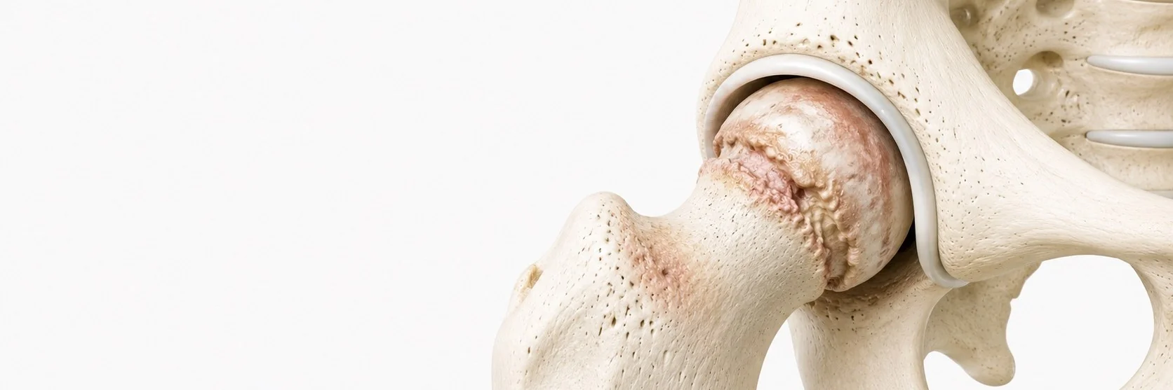
What is hip osteoarthritis?
Hip osteoarthritis is a progressive degenerative condition of the hip joint, where the smooth articular cartilage covering the femoral head and the acetabulum gradually wears away. As cartilage is lost, bone-on-bone contact develops, leading to pain, stiffness and progressive loss of function.
It is one of the most common causes of disability in older adults, but it can also affect younger patients with predisposing anatomy or previous injury.
What causes it?
Hip osteoarthritis can be:
- primary, without an obvious underlying cause, related to age, genetics and biomechanical factors
- secondary, following a specific cause such as femoroacetabular impingement, hip dysplasia, previous trauma, avascular necrosis, or inflammatory arthritis
Identifying secondary causes is important, it can lead to earlier intervention and, in younger patients, joint-preserving treatment options.
What are the symptoms?
- groin pain, the most characteristic symptom; pain may also radiate to the thigh or buttock
- pain that worsens with activity, particularly walking and stairs
- stiffness, especially in the morning or after sitting for long periods
- difficulty putting on socks and shoes (a classic early sign)
- limp, particularly after prolonged walking
- reduced range of motion, especially internal rotation
- in advanced disease, rest pain and night pain
How is the diagnosis made?
- clinical examination, assessing range of motion, gait, leg length and pain pattern
- X-rays, the cornerstone of diagnosis, showing joint space narrowing, osteophytes, sclerosis and cysts
- MRI, used selectively for early disease, to evaluate cartilage and exclude avascular necrosis
X-ray staging guides treatment recommendations and helps set realistic expectations for outcomes.
What is the conservative treatment?
For mild to moderate disease, non-surgical treatment is the starting point:
- weight management, reduces load on the joint substantially
- low-impact exercise, swimming, cycling, walking on level ground
- physiotherapy focused on hip mobility, gluteal strength and gait
- analgesics and anti-inflammatory medication when appropriate
- walking aids in selected cases
- Molecular & Biological Regenerative Therapy (PRP) for early disease
Conservative measures help symptoms but do not reverse cartilage loss. As disease progresses, their effect typically diminishes.
When is hip replacement considered?
Total hip replacement is considered when:
- pain significantly limits daily activities, work or sleep
- walking distance is reduced
- X-rays show advanced cartilage loss
- conservative treatment has been optimised but is no longer adequate
How is hip replacement performed?
Modern total hip replacement is one of the most successful operations in medicine. The worn femoral head and acetabular cartilage are replaced with prosthetic components, typically a metal stem, a ceramic head and a polyethylene liner in a metal cup.
I use modern bearings and minimally invasive approaches when appropriate, aiming to preserve soft tissues and accelerate recovery. The implant choice is individualised based on the patient's anatomy, age and activity level.
What should the patient expect after surgery?
- most patients walk on the day of surgery with crutches
- hospital stay is typically 2–4 days
- crutches are gradually discontinued over 2–6 weeks
- driving usually resumes at 4–6 weeks
- return to office work at 4–6 weeks; more demanding work at 3 months
- return to recreational activities (cycling, swimming, golf) at 3 months
- physiotherapy is essential and structured, early mobilisation, then progressive strengthening
What is the long-term outcome?
Total hip replacement has excellent and predictable outcomes. Patient satisfaction routinely exceeds 90%. Modern implants have 15-year survivorship over 95% in national registries, and many last 25 years or more.