

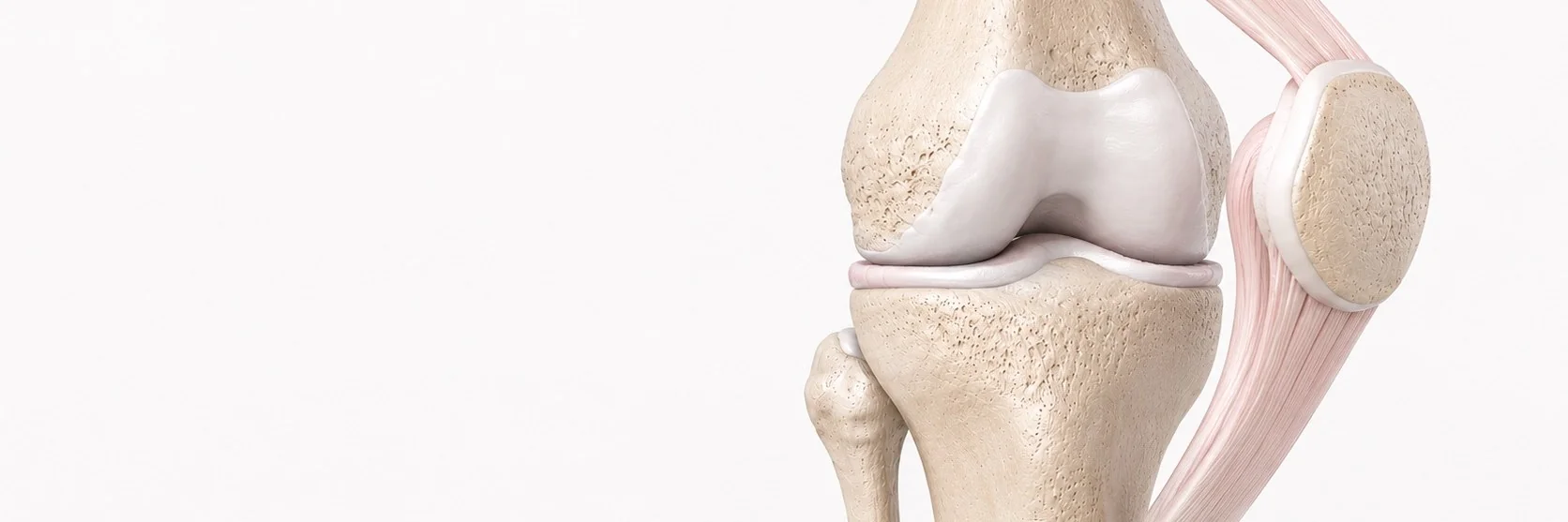
What is patellar instability?
Patellar instability is a condition in which the patella (kneecap) moves abnormally outside its normal position within the trochlear groove of the femur. It can range from a single traumatic dislocation to chronic recurrent instability that interferes with daily life and sport.
What causes patellar instability?
Patellar instability is usually multifactorial. The main contributing factors are:
- trochlear dysplasia, abnormal shape of the trochlear groove
- patella alta, a high-riding patella that engages the groove later in flexion
- increased TT-TG distance, abnormal tracking of the extensor mechanism
- MPFL (medial patellofemoral ligament) deficiency, the primary medial restraint of the patella, often torn after a dislocation
- generalised ligamentous laxity
- valgus malalignment of the lower limb
What are the symptoms?
- a frank dislocation event, often dramatic and painful
- a feeling of the patella "slipping" or giving way with twisting movements
- anterior knee pain
- swelling after instability episodes
- apprehension and avoidance of certain movements
- in chronic cases, secondary cartilage damage and progressive patellofemoral arthritis
How is the diagnosis made?
- clinical history and examination, including the apprehension test
- X-rays, to assess patellar height, trochlear shape and overall alignment
- MRI, for evaluation of the MPFL, articular cartilage and trochlear morphology
- CT scan with measurement of TT-TG distance and assessment of trochlear dysplasia, particularly when surgery is being planned
What is the conservative treatment?
After a first dislocation in a patient without significant anatomical risk factors, conservative treatment is usually appropriate:
- short-term immobilisation followed by progressive mobilisation
- structured physiotherapy with emphasis on quadriceps (especially VMO), hip and core strengthening
- patellar taping or bracing during return to activity
- addressing biomechanical factors
When is surgery recommended?
Surgery is recommended when:
- there are recurrent dislocations despite a properly conducted rehabilitation programme
- there is a clear anatomical risk factor (significant trochlear dysplasia, patella alta, increased TT-TG)
- the first dislocation has caused a major chondral or osteochondral injury that itself requires surgery
- there is significant ongoing functional limitation
How is surgery performed?
Patellar instability surgery must address all the underlying anatomical factors that have made the patella unstable. There is no single operation that fits every patient. The procedures I use, alone or in combination, include:
- MPFL reconstruction, the cornerstone procedure for most cases, using a tendon graft to recreate the medial restraint of the patella
- tibial tubercle osteotomy, to correct an abnormal TT-TG distance and/or patellar height
- trochleoplasty, for severe trochlear dysplasia (a complex procedure reserved for selected cases)
- treatment of any associated cartilage damage
Dynamic MPFL reconstruction
I perform modern anatomic MPFL reconstruction using a tendon autograft, with careful femoral and patellar fixation under fluoroscopic control to avoid over- or under-tensioning the new ligament. When indicated, I combine MPFL reconstruction with a distalising or medialising tibial tubercle osteotomy to address the underlying biomechanical problem. I apply modern techniques aiming for stable repair and optimal functional recovery.
What should the patient expect after surgery?
- knee brace for 4–6 weeks
- early protected range-of-motion
- weight-bearing as tolerated after isolated MPFL; protected weight-bearing for 6 weeks after osteotomy
- structured physiotherapy with progressive strengthening
- return to sport at 4–6 months for isolated MPFL, 6–9 months when combined with osteotomy