

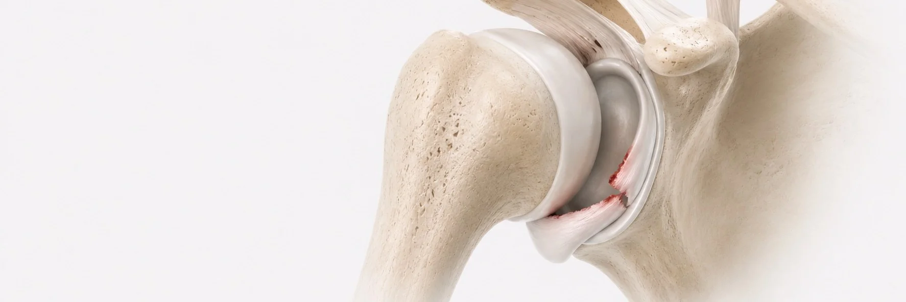
What is shoulder instability?
The shoulder is the most mobile joint in the human body, and that mobility comes at the price of stability. Shoulder instability is the condition in which the head of the humerus slips partially (subluxation) or completely (dislocation) out of the glenoid socket. It can range from a single traumatic dislocation in a young athlete to chronic recurrent instability that interferes with daily life.
There are three main patterns:
- anterior instability, by far the most common (around 95% of cases), usually after a traumatic dislocation
- posterior instability, less common, often related to seizures, repetitive overhead loading or contact sports
- multidirectional instability (MDI), instability in more than one direction, frequently associated with generalised ligamentous laxity
What are the causes?
- traumatic, a fall on an outstretched arm, a tackle in contact sports, a forced overhead movement
- atraumatic / repetitive, chronic overuse in throwers, swimmers, gymnasts; gradual stretching of the joint capsule
- constitutional laxity, naturally lax connective tissue (Ehlers-Danlos and similar syndromes), more common in young women
- previous dislocation, the strongest single risk factor for further instability is a first dislocation, particularly under the age of 25
What structures are damaged?
A traumatic anterior dislocation typically produces a combination of injuries:
- Bankart lesion, detachment of the anterior labrum from the glenoid
- Hill-Sachs lesion, an impression fracture on the back of the humeral head
- bony Bankart, fracture of the front of the glenoid rim (in higher-energy injuries)
- capsular and ligamentous stretching
- less commonly, rotator cuff or nerve injuries (especially in older patients)
What are the symptoms?
- a frank dislocation event, often dramatic and painful, sometimes requiring reduction in the emergency department
- a sense of the shoulder "slipping out" with specific movements (apprehension)
- recurrent episodes of instability with progressively less force required
- pain, weakness and reduced sporting performance
- anxiety and avoidance of certain positions or activities
How is the diagnosis made?
- clinical examination with apprehension and relocation tests
- X-rays to assess the position of the humeral head and to identify bony lesions
- MRI arthrogram, the most sensitive imaging modality for labral and capsular injuries
- CT scan, essential when there is significant bone loss on the glenoid or humeral head, because the amount of bone loss directly determines the type of surgery required
What is the conservative treatment?
After a first traumatic dislocation, treatment includes:
- closed reduction of the dislocation
- short-term immobilisation in a sling
- a structured physiotherapy programme focused on rotator cuff and scapular stabiliser strengthening, proprioception and dynamic control
For atraumatic or multidirectional instability, conservative treatment is the first-line management for at least six months, because surgery in this group is less reliable. In my experience, a well-conducted rehabilitation programme is essential before considering surgery.
When is surgery recommended?
Surgery is offered when:
- recurrent instability persists despite an adequate rehabilitation programme
- there is significant bony injury (large Hill-Sachs or glenoid bone loss greater than 15–20%)
- the patient has high functional demands (young athletes, contact-sport players) where the recurrence rate without surgery is very high
- instability is significantly affecting work, sport or daily life
How is surgical treatment performed?
The surgical strategy depends on what is damaged and how much bone is lost.
Soft-tissue procedures (most cases):
- arthroscopic Bankart repair, the gold standard for typical anterior instability without significant bone loss. The torn labrum is reattached to the glenoid with anchors, restoring the bumper that prevents the humeral head from slipping forwards
For shoulders with significant glenoid bone loss, an open bone-augmenting operation (such as Latarjet) is the most reliable approach, but I do not perform this procedure myself. In these cases I refer the patient onward to a specialist colleague after a clear discussion of the indication and the rationale.
Posterior and multidirectional instability are typically treated with arthroscopic capsulolabral repair and capsular plication.
I apply modern arthroscopic techniques aiming for stable repair and optimal functional recovery.
What should the patient expect after surgery?
After arthroscopic Bankart repair:
- sling for 4–6 weeks
- passive range of motion in weeks 1–6, progressive active motion in weeks 6–12
- strengthening from 3 months
- return to non-contact sport at 4–5 months, contact sport at 6 months
- recurrence rate below 10% in correctly selected patients
The choice of procedure is individualised, there is no single "best" operation for every patient. The decision is based on bone quality, sport, expectations and previous surgery, and is made together with the patient after detailed discussion.